Low Vitamin D in infants - video interview of Dr. Holick
Video on YouTube
A few notes of the video
Ehlers-Danlos syndrome (EDS) 1 in 5,000 people
EDS results in hypermobility of joints and skin
If one parent has EDS then 50% chance that the infant will have EDS
If two parents have EDS then 75% chance in infant
No DNA test (yet) for EDS
EDS very dangerous if also have low vitamin D. Looks like broken bones due to child abuse
EDS on Wikipedia 13 types of EDS as of 2017
Best source of Calcium Milk or yogurt (especially if lactose intolerant - also TUMS)
No need to test, very low cost, just supplement
If mother minimally took prenatal Vitamins (400 IU) highly likely that both mother and infant will be deficient
Legs can be bowed out or bowed in with Rickets
Head sweating in infant is a good sign of low vitamin D
Infant can bruise easily with rickets
Appears to be discussing his Sept 2016 study
Multiple unexplained fractures in infants and child physical abuse.
J Steroid Biochem Mol Biol. 2016 Sep 15. pii: S0960-0760(16)30248-5. doi: 10.1016/j.jsbmb.2016.09.012
Cannell JJ1, Holick MF2.
When an infant presents with X-rays showing multiple unexplained fractures in various stages of healing (MUFVSH), the child is usually diagnosed with child abuse based on criteria of the Academy of Pediatrics' Committee on Child Abuse and Neglect (AAPCCAAN). Almost always, the infant is subsequently removed from the home and civil or criminal proceeding commence. It may be that healing infantile rickets or other poorly understood metabolic bone disorders of infancy are responsible for these x-rays. Activated vitamin D is a seco-steroid hormone, whose mechanism of action is genetic regulation. Lack of it can result in musculoskeletal defects known as rickets. Low calcium can also cause rickets. However, it is clear that experts for the state believe that the x-rays in these cases are so definitive as to be pathognomonic for child abuse. Therefore, if the caregivers deny abusing their infants, experts following American Academy of Pediatric's Committee on Child Abuse and Neglect. guidelines are essentially claiming that x-rays showing multiple unexplained fractures in various stages of healing are lie detector tests. However, it is not widely appreciated that the gold standard for the diagnosis of rickets is a bone biopsy, not x-rays, as radiologists miss biopsy proven rickets 80% of the time; that is, 4 out of 5 infants with rickets will have normal x-rays . In this article we provide reports of two cases and their outcomes. We discuss information about healing infantile rickets and an example of common sense medical conclusions in these cases. This information could lead to a significant reduction in the number of innocent parents having their infant removed or sent to prison.
📄 Download the PDF from SciHub via Vitamin D Life
Audio-only interview April 2017
What is vitamin D
Is it possible to reduce the risk of the flu by as much as 42%
Vitamin D controls over 200 genes in the cardiovascular system
Reduce the risk of heart attack by 50%
Lack of Vitamin D causes higher blood pressure
Why we who live in Norway have 10-15x greater likelihood of diabetes
How much vitamin D your body needs every single day
What are the symptoms of too little D vitamin
Why is vitamin D so incredibly important to us
What diseases can you get because of little vitamin D
How long do you have to be in the sun to limit the likelihood of high blood pressure and illnesses?
Do you live in Norway? Your body does NOT produce vitamin D 6 months of the year.
If are you born in the winter, you have a higher risk of Schizophrenia later in life.
Higher vitamin D values reduce the risk of cesarean section by 200 to 300%
How high values should you have while you are pregnant
How much D Vitamin should have small children every day
Why your skin turns red when you're in the sun? Use of sunscreen
The most dangerous form of skin cancer occurs when you do not get sun.
JAMA wondered about EDS - Oct 2017
Flawed Theories to Explain Child Physical Abuse: What Are the Medical-Legal Consequences?
📄 Download the PDF from Sci-Hub via Vitamin D Life
Cited 34 times as of June 2024 - appears to both agree and disagree
Vitamin D deficiency in patients with Ehlers–Danlos syndrome - 2024
Endocrine Abstracts (2024) 99 EP545 | DOI: 10.1530/endoabs.99.EP545
Poprawa Iga 1,2 , Babula Emilia 1,2 , Żuk-Łapan Aleksandra 1,2 , Podstawka Jakub 1,2 , Julia Latocha 1,2 , Zuzanna Szymańska 1,2 , Domański Jan 1,2 , Bernadetta Kaluza 1,2 & Franek Edward 1,2,3
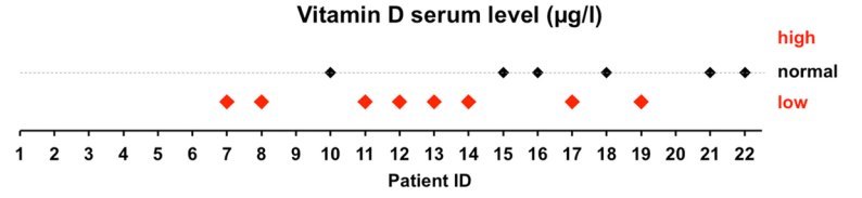
Background : Ehlers–Danlos syndrome encompasses a group of genetic conditions characterized by alterations in connective tissue structure, with the consequent increased risk of developing osteopenia, osteoporosis, and incurring fractures. Maintaining vitamin D levels within normal limits is known to play an important role in preventing these complications, which seems to be particularly important in patients with Ehlers–Danlos syndrome. The purpose of this study was to assess serum 25-hydroxyvitamin D, or 25(OH)D, levels in women with Ehlers–Danlos syndrome.
Material and methods: The study involved a prospective assessment of 30 female patients, aged 20–53 years, with hypermobile or classical Ehlers–Danlos syndrome. All patients underwent calcium and phosphorus metabolism testing and bone mineral density (BMD) scans of the femoral neck and lumbar spine. The patients were divided into two groups: those with vitamin D deficiency, defined as serum 25(OH)D levels of < 30 ng/ml (group 1, n=18) and those with normal (> 30 ng/ml) 25(OH)D levels (group 2, n=12).
Results: Eighteen patients (60%) showed vitamin 25(OH)D deficiency, with three of those (16.7%) showing secondary hyperparathyroidism. Study groups 1 and 2 showed no significant differences in terms of serum levels of calcium (2.4±0.09 mmol/l vs 2.39±0.07 mmol/l, P=0.88), phosphorus (3.51±0.7 mg/dl vs 3.42±0.51 mg/dl, P=0.86), bone-specific alkaline phosphatase (10.36±3.06 µg/l vs 8.28±1.31 µg/l, P=0.007), beta-CrossLaps (0.39±0.19 ng/ml vs 0.39±0.17 ng/ml, P=0.69), or osteocalcin (20.14±8 ng/ml vs 21.23±6.67 ng/ml, P=0.46), femoral neck BMD (0.95±0.12 g/cm2 vs 0.92±0.13 g/cm2, P=0.57), or lumbar spine BMD (0.12±0.16 g/cm2 vs 0.13±0.11 g/cm2, P=0.14).
Conclusions: Sixty percent of patients with Ehlers–Danlos syndrome showed vitamin 25(OH)D deficiency . Parameters of calcium–phosphorus metabolism in these patients were not significantly different from those in patients with normal serum vitamin 25(OH)D levels.
Vascular type Ehlers-Danlos syndrome is associated with platelet dysfunction and low vitamin D serum concentration - 2016
📄 Download the PDF from Vitamin D Life
Shaken Baby Syndrome, etc. in Vitamin D Life
Hospital has banned Dr. Holick due to testifying in cases of infants with broken bones- August, 2021
Child abuse fractures – 96 percent were associated with poor bones (low vitamin D, etc.) – Oct 2019
The Vitamin Deficiency Signs That Can Send You to Prison – Feb 2014
Shaken Baby Syndrome - probably caused by Ehlers-Danlos Syndrome
Lack of vitamin D in infants can result in broken bones and shaken baby syndrome - March 2010
Child abuse, vitamin D deficiency, or what - for parents and defense attorneys - Cannell June 2015
Mother and father on trial for infant death – set free – death was due to rickets – Dec 2011
Childhood Fractures – consensus report on vitamin D – Feb 2016
Some Rickets is due to poor genes - Vitamin D needed lifelong – June 2020
Shaken Baby Syndrome - Trailer April 2016
55+ Vitamin D Life pages have HOLICK in the title
{LIST()}